
Guides
Advanced MRI
A larger Siemens study with the series that need care: what the scan leaves out and why, maps the scanner computes from a diffusion run, a sequence named one thing and acquired as another, and spectroscopy with its water reference.
The sample
Three visits on a Siemens Prisma: OL_3844 and OL_3845 are one participant on two days, OL_3846 another. Between them: T1-weighted, T2-weighted and FLAIR images, gradient-echo images made for neuromelanin, BOLD runs with their references and physiological logs, two diffusion runs with reversed-phase references and the maps the scanner computed, fieldmaps, and single-voxel spectroscopy with its water reference.
What the scan leaves out
Step 1. Scan and read the left-out rows
Create a dataset named mri_advanced_study and scan the sample folder: 51 rows in 47.3 seconds, 31 of them to convert.
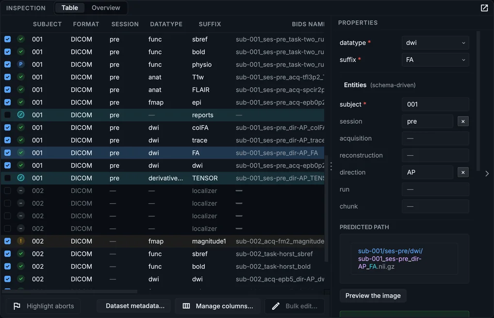
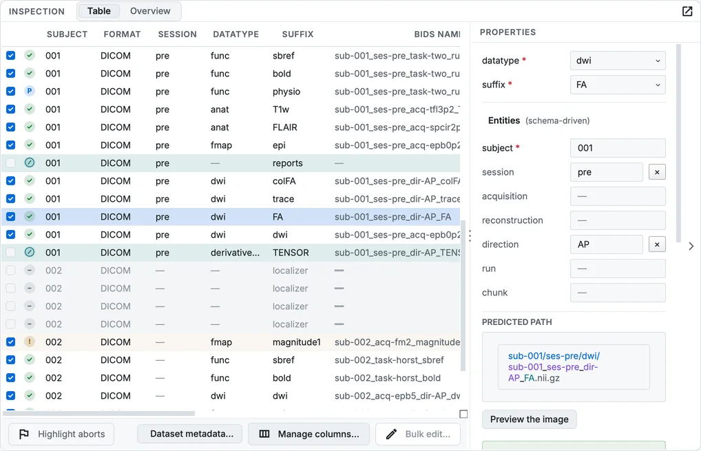
The scan, scrolled to the diffusion series.
| Left out | Why |
|---|---|
AAHead_Scout_… (several per visit) |
Scout images for positioning the slices. |
PhoenixZIPReport |
The scanner's protocol report: DICOM, but no image. |
ses-pre_dir-ap_dwi_TENSOR |
The diffusion tensor the scanner computed: stored without an image, so nothing can be converted. |
Kept, although the scanner computed them: …_dwi_FA, …_dwi_ColFA and …_dwi_TRACEW. BIDS has names for these maps in dwi/ (FA, colFA, trace), so they are converted beside the diffusion run. Untick them if your analysis recomputes them, as most do.
A series named one thing and acquired as another
The series ses-post_ was named T1w by the operator. dcm2niix's guess, from its header strings, is T2starw. The parameter classifier reads how it was acquired and agrees with the operator, and Why this name shows why:
Step 2. Read the evidence
Select the row and scroll Properties to Why this name.
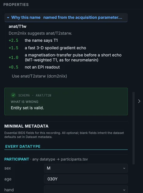
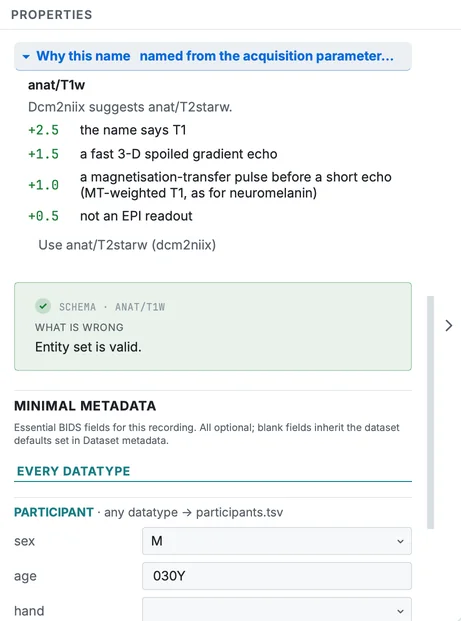
Why this name for the neuromelanin gradient echo.
A fast 3-D spoiled gradient echo with a magnetisation-transfer pulse before a short echo is a T1-weighted image of the kind made for neuromelanin. The classifier named it anat/T1w with part-mag, and listed dcm2niix's T2starw as the alternative. If your protocol calls it something else, change the suffix in the table.
The same classifier names ses-pre_T2w, a SPACE sequence with an inversion pulse, FLAIR: the course explains that case (Naming recordings).
Spectroscopy
OL_3846 holds two spectroscopy series: the metabolite spectrum and its water reference, both proposed as svs runs 1 and 2. The reference is not a second run: select its row (its series name contains type-ref), set suffix to mrsref and clear run; clear run on the spectrum too. The course does the same on its sample (Naming recordings). After the conversion, the spectrum opens in the Editor as a spectrum (Viewing data).
Convert and check
Step 3. Convert
Give the authors and licence in Dataset metadata… and click Run conversion. The sample converted in 23.9 seconds into 111 files; the second diffusion run alone is 8,775 DICOM files.
Step 4. Look at the diffusion folder
In the Editor, open sub-001/ses-pre/dwi/. The run's .bval opens as its gradient table; the scanner's maps are files of their own beside it.
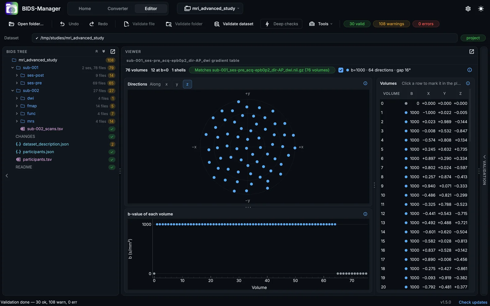
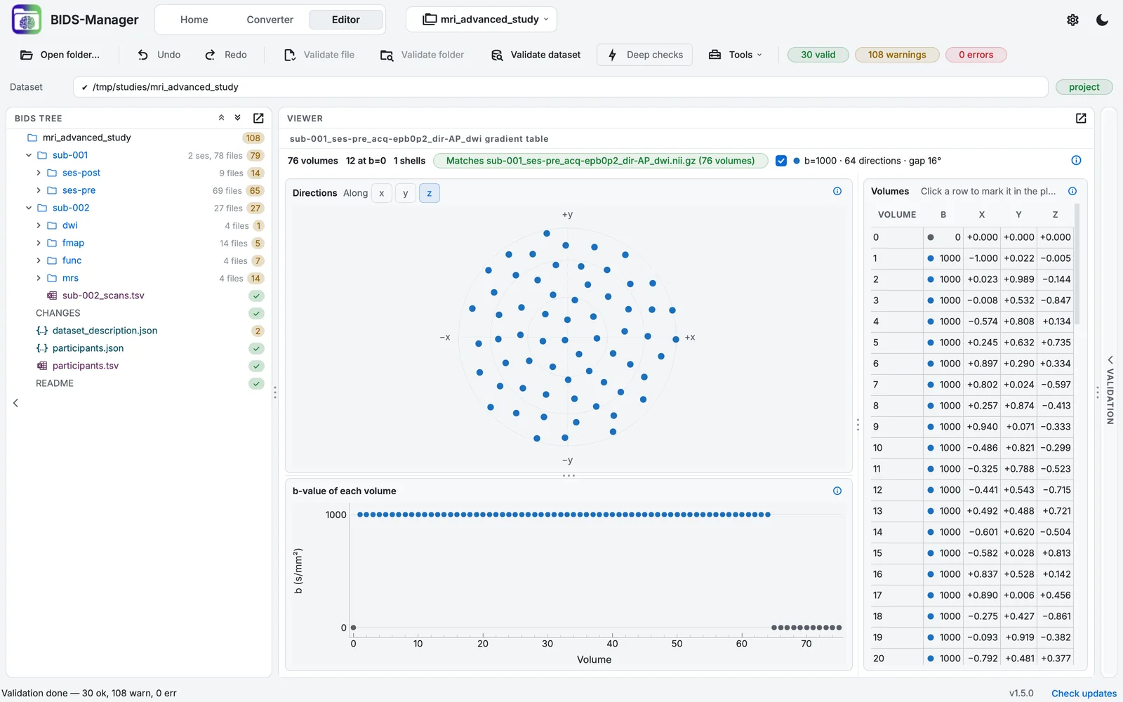
The diffusion folder of session pre.
Validate dataset reports 30 files valid, 108 warnings and 0 errors.
Identifiers and faces
The participants of this sample consented to their faces being shown. For your own data: the conversion removes the participant's identifiers from every sidecar and from the spectroscopy files; check participants.tsv, deface the anatomical images, and do not share sourcedata/ (Identifiers and privacy).