
Learn · Chapter 13 of 15
Checking image quality
Run the fast quality check on an anatomical image, read the plots under a BOLD run, check every image of the dataset at once, and look at an MEG recording's channels. The checks run on your computer in seconds and change nothing in the data.
What the quality check is
The quality check measures what can be measured on one image without a full processing pipeline: noise and contrast, artefacts in the air around the head, how the tissues separate, coverage and smoothness, and for diffusion the gradient table, motion and slice dropouts. It follows the definitions of MRIQC where they apply, and every measure is explained where it is shown (hover it, or click its info icon) and in Quality measures.
It is for screening: an image that looks wrong in the numbers is worth looking at. It does not replace looking at the images.
Check an anatomical image
Step 1. Open the T1w and check it
Select the T1w in the Editor, then choose QC > Check image quality in the viewer's toolbar. The quality panel opens beside the planes; the check takes about half a minute on the sample's T1w. The first check on a computer also downloads the two brain models it uses, about 1.5 MB.
The quality check of the T1w beside the Planes + 3-D layout, laying its masks and tissue classes over the image.
Step 2. Read the panel
From the top:
- what was checked and how: the brain was found with
mindgrab, the tissues withrobust_tissue, the registration withniimath; - On the image: the masks and maps the check made, each a checkbox that lays it over the image (brain mask, head mask, the air measured, CSF, grey and white matter, the bias field, the face region);
- the findings, each with Show: on the sample, A face is visible, because the sidecar records no defacing and the face region holds tissue. Chapter 14 deals with it;
- the Measures, grouped (noise and contrast, artefacts, tissues, coverage and smoothness), each with its value and its explanation.
The air around the head is measured only in images that are not defaced, not skull-stripped and not set to zero by the scanner, because in those it says nothing about noise.
The QC plots of a BOLD run
Select the BOLD run, turn on Time course, and tick QC in its header. Plots chooses which plots appear under the time course, aligned with it in time:
The BOLD run's QC plots under its time course, following the run as it plays.
| Plot | What it shows |
|---|---|
| Framewise displacement | How far the head moved from one volume to the next, in millimetres |
| Translation, Rotation | The head's position over the run |
| DVARS | How much the whole image changed from one volume to the next |
| Outlier voxels | The share of voxels far from their usual value, per volume |
| Slice spikes | A slice whose signal jumps away from its own course in one volume, brighter or darker |
| Global signal | The mean over the head, per volume |
| Carpet plot | Every voxel's time course as a row, so a sudden change shows as a vertical band |
QC in the viewer's toolbar also offers maps of the whole run: Temporal SNR, Standard deviation and Mean image.
Check every image at once
Tools > Quality check... checks every anatomical and diffusion image of the dataset and lists them with their findings and main measures. Click Check; At once sets how many images are checked in parallel.
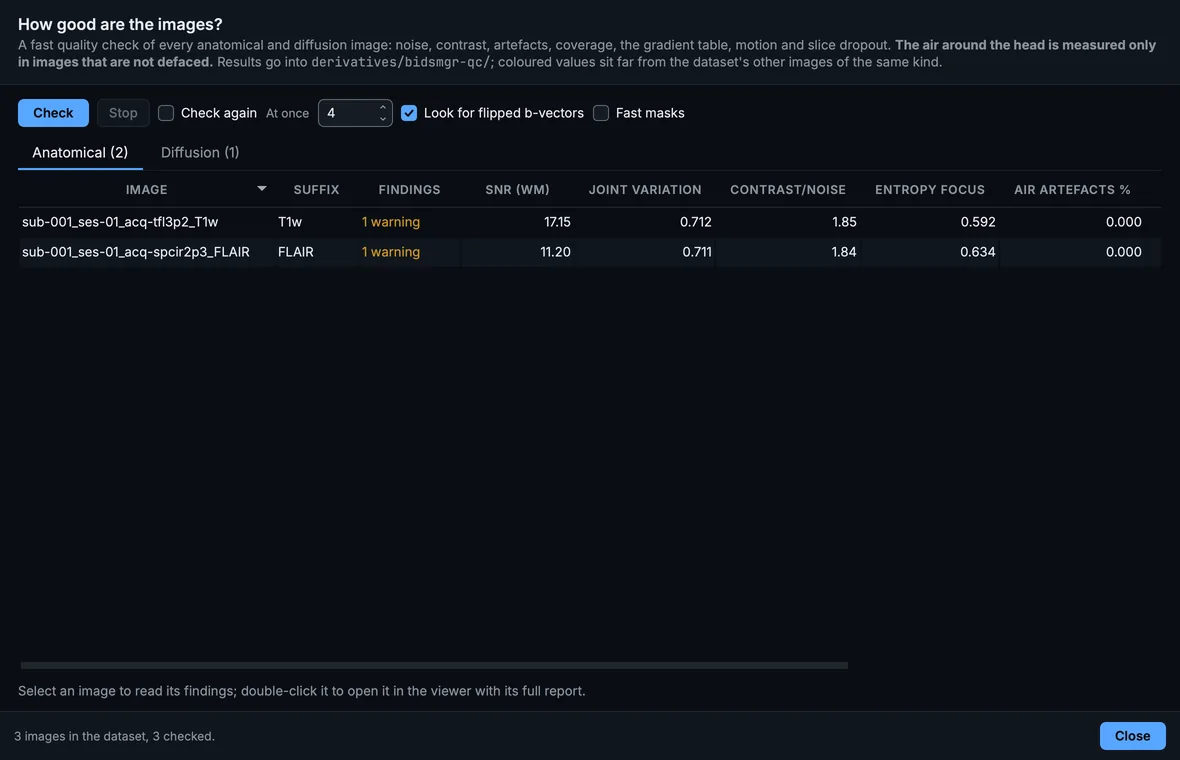
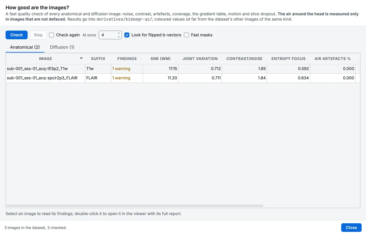
The quality check of the whole dataset.
Double-click an image to open it in the viewer with its full report. The results are saved in derivatives/bidsmgr-qc/ in the dataset, so they are not computed twice. The conversion can run the same check at its end (Settings > Convert > Check image quality).
MEG and EEG
In the signal viewer, QC checks the recording channel by channel, per segment: channels much noisier or flatter than the others of their type, line noise, segments where many channels jump, and muscle activity. Plots chooses what is drawn beside the traces; Mark as bad turns a finding into a bad channel or segment, which Save to dataset writes.
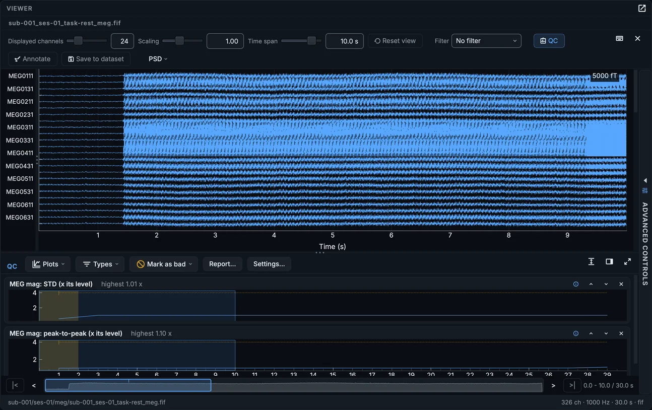
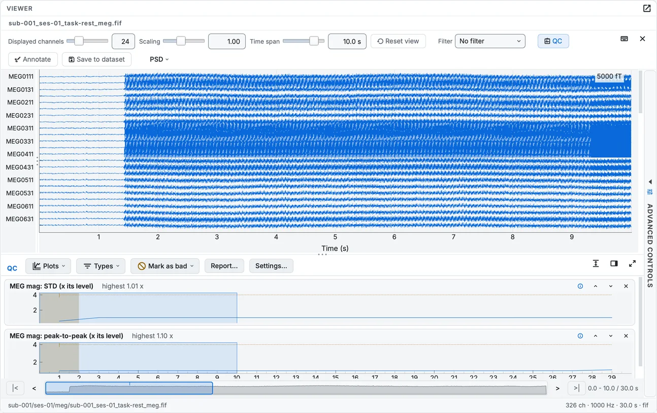
The quality check of the resting MEG recording.
Settings
Settings > Quality control holds when QC runs (Run QC when a file opens), the methods (brain mask, tissue classes, registration), and the thresholds of every check, each with its range. The values are in Settings.
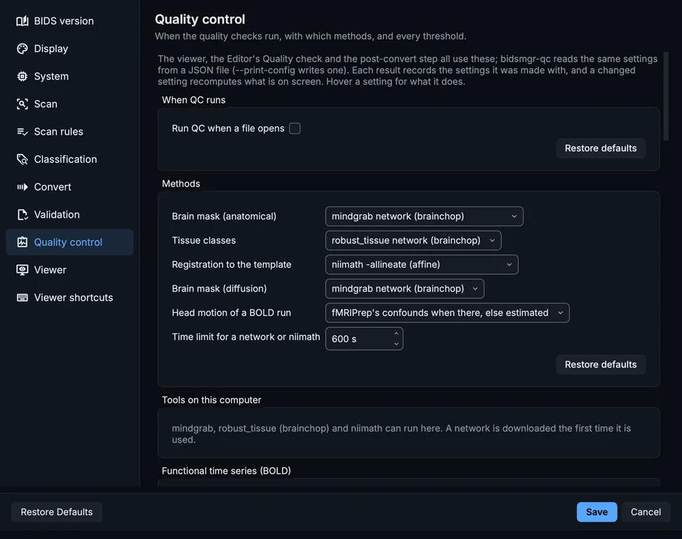
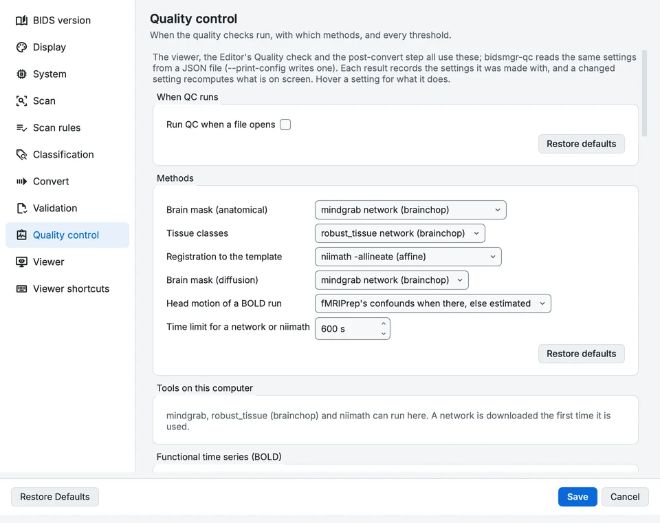
Settings, Quality control page.
Your progress is kept in this browser only.